Understanding Digital X-Ray Imaging
What is Digital X-Ray Imaging?
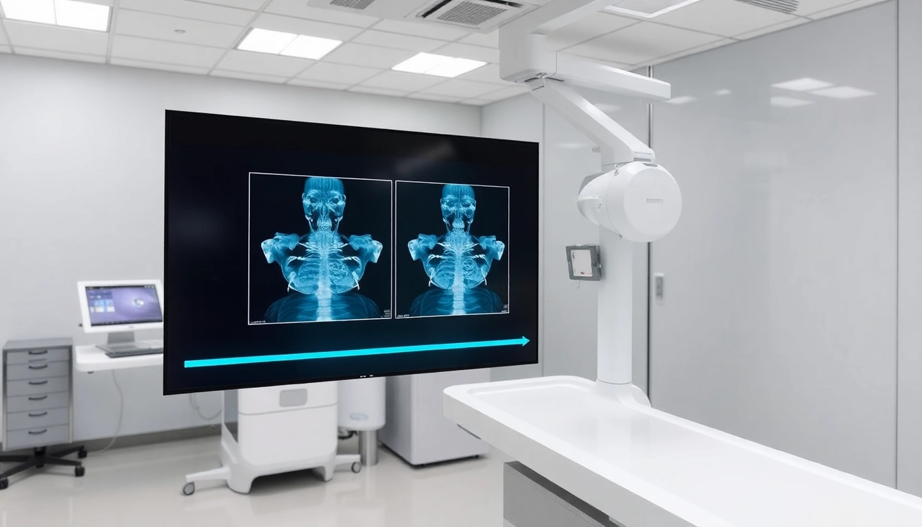
Digital X-ray imaging represents a significant leap forward in medical imaging technology, utilizing digital detectors instead of traditional film to create images of the internal structures of the body. This innovative approach generates images that can be processed, viewed, and stored electronically, offering numerous advantages over conventional methods. As healthcare continues to evolve, understanding digital X-ray imaging becomes increasingly essential, with its impact on patient care, accuracy in diagnosis, and overall healthcare efficiency becoming ever more profound. Within this landscape, the future of digital X-ray imaging is poised for remarkable developments, driven by ongoing technological advancements.
Comparison of Traditional vs. Digital X-Ray Techniques
Traditional X-ray imaging, which relies on the use of film to capture images, has been the cornerstone of radiological practices for decades. However, the transition to digital X-ray techniques has radically transformed how medical imaging is conducted. Key differences include:
- Image Quality: Digital images offer enhanced resolution and clarity, allowing for better visualization of subtle details compared to traditional film.
- Speed: Digital X-rays can be processed almost instantaneously, enabling faster diagnosis and treatment decisions.
- Storage and Accessibility: Digital images can be easily stored in electronic health records, facilitating rapid access by multiple healthcare providers.
- Environmentally Friendly: The elimination of film and chemicals required in traditional X-ray processes reduces environmental waste and improves safety for technicians.
Key Advantages of Digital X-Ray Imaging
The shift from traditional to digital X-ray imaging is underpinned by several notable advantages that cater to both healthcare providers and patients:
- Enhanced Diagnostic Accuracy: With features like contrast adjustment and image enhancement, digital images can be modified to reveal even the faintest abnormalities, thereby increasing diagnostic accuracy.
- Reduced Radiation Exposure: Digital X-rays require lower doses of radiation compared to film X-rays without compromising image quality, making them safer for patients.
- Cost-Effectiveness: Although the initial investment in digital systems can be significant, the long-term savings related to film, processing costs, and physical storage make them a wise financial choice.
- Improved Patient Workflow: Patients benefit from shorter wait times and a more streamlined diagnostic process, leading to improved satisfaction and outcomes.
Technological Advancements in Digital X-Ray Imaging
Emerging Technologies in Digital X-Ray
The landscape of digital X-ray imaging is continuously evolving, with several emerging technologies making significant impacts. These include:
- Flat Panel Detectors: These devices are becoming the standard in digital radiography, providing high-quality images with faster acquisition times and improved sensitivity.
- 3D Imaging Techniques: Innovations in three-dimensional imaging allow for comprehensive visualization of complex anatomical structures, facilitating better diagnosis and pre-surgical planning.
- Wireless Imaging Systems: The advent of wireless systems enhances mobility within medical facilities, allowing imaging to be performed anywhere with immediate availability of results.
- Image Fusion Technologies: Combining X-ray images with other imaging modalities, such as CT or MRI, helps in creating a more complete picture of a patient’s condition.
Integration of AI in X-Ray Imaging
Artificial Intelligence (AI) is revolutionizing the field of medical imaging, particularly in digital X-ray. AI algorithms are being developed to assist radiologists by:
- Automated Detection: AI can quickly analyze images, identifying abnormalities that may be missed by the human eye, thereby reducing diagnostic errors.
- Predictive Analytics: By correlating imaging features with patient data, AI can predict potential health issues, leading to proactive patient management.
- Workflow Optimization: AI systems can streamline workflows by prioritizing cases that require urgent attention, improving efficiency within radiology departments.
Potential Future Developments in Imaging Tech
The future of digital X-ray imaging holds exciting possibilities as various technological advancements continue to reshape the industry. Potential developments include:
- Enhanced Image Processing: Future algorithms will be able to provide real-time image analysis, further enhancing diagnostic accuracy and reducing the time to interpret results.
- Tele-radiology Advances: As telehealth continues to expand, advancements in remote imaging technologies will facilitate instant sharing of X-ray images for consultation across geographical boundaries.
- Integration of Virtual Reality: Virtual reality could enable more interactive training for radiologists, improving diagnostic skills and enhancing patient education.
- Personalized Imaging Techniques: Tailoring imaging modalities to individual patient needs based on genetic, behavioral, and environmental factors may optimize diagnostic approaches.
Impact on Patient Care and Diagnosis
Improving Accuracy and Speed of Diagnosis
The implementation of digital X-ray imaging significantly boosts the accuracy and speed of diagnosis. By facilitating quicker access to clearer images, radiologists are better positioned to make timely and accurate diagnoses. For instance, studies have shown that digital X-rays lead to a reduction in misdiagnosis rates, ultimately improving patient outcomes. Rapid image processing allows for immediate assessments, often resulting in faster treatment plans that are critical in emergency situations.
Enhancing Patient Experience with Digital Imaging
Digital X-ray imaging not only improves diagnostic accuracy but also enhances the overall patient experience. Patients benefit from less radiation exposure, quicker procedures, and more transparency in their healthcare journey. The reduction in wait times for imaging results fosters trust and satisfaction, which is essential for patient retention and positive health outcomes. Moreover, digital systems often allow for easier sharing of results with patients, enabling them to engage actively in their care.
Real-World Case Studies of Success
There are numerous real-world examples that illustrate the successful implementation of digital X-ray imaging systems in various healthcare settings:
- Hospital A: In a metropolitan hospital that transitioned to a fully digital X-ray system, diagnostics for fractures were completed 30% faster, leading to improved patient throughput and higher satisfaction ratings.
- Clinic B: A small orthopedic clinic adopted digital imaging and saw a 25% reduction in follow-up appointments due to increased diagnostic precision, enhancing their operational efficiency.
- Radiology Center C: After incorporating AI-driven diagnostic tools, this center reduced the time for image interpretation by 40%, enabling a more rapid response to patient needs.
Challenges and Considerations
Common Misconceptions about Digital X-Ray Imaging
Despite the numerous advantages of digital X-ray imaging, several misconceptions persist, such as:
- Cost Implications: Many believe that transitioning to digital systems is prohibitively expensive, overlooking the long-term savings on film, processing, and storage.
- Training Requirements: Some assume staff will require extensive training for digital systems, whereas most modern systems are designed for user-friendly operation with minimal training.
- Image Quality Concerns: There is a misconception that digital images may be inferior to traditional images; however, advancements continuously debunk this belief with enhanced imaging techniques.
Addressing Data Privacy and Security Issues
As digital X-ray imaging moves toward greater integration with electronic health records, the issue of data privacy and security becomes paramount. Effective measures must be implemented to protect patient information from breaches. Healthcare providers should consider the following best practices:
- Encryption: Utilizing encryption for data storage and transmission can protect sensitive information from unauthorized access.
- Access Controls: Implementing strict access controls and permissions ensures that only authorized personnel can view and manage imaging data.
- Regular Training: Staff should receive training on best practices for maintaining data security and confidentiality compliance.
Cost Implications of Transitioning to Digital Systems
Transitioning to digital X-ray imaging involves initial setup costs that can be substantial. However, it is vital to consider the total cost of ownership, including:
- Reduced Operating Costs: Digital imaging eliminates costs associated with film, chemicals, and processing, leading to savings over time.
- Increased Efficiency: Improved workflows can translate to greater patient throughput, ultimately increasing revenue opportunities.
- Long-term ROI: While the upfront investment may seem daunting, the long-term return on investment benefits outweigh the initial costs as streamlined operations and reduced waste enhance profitability.
Best Practices for Implementing Digital X-Ray Solutions
Steps for Transitioning to Digital X-Ray Technology
The transition to digital X-ray technology can be streamlined by following key steps:
- Assessment of Needs: Evaluate current systems and identify specific needs that digital X-ray technology must address.
- Budget Planning: Create a detailed budget that encompasses not only the initial purchase but also ongoing maintenance, training, and operational costs.
- Vendor Selection: Research and select reputable suppliers who provide quality products and strong customer support.
- Implementation Timeline: Develop a realistic timeline for transition to minimize disruption to clinical services.
Training Staff for New Digital Systems
Proper training is essential for maximizing the benefits of digital X-ray technology. Best practices for training include:
- Hands-On Training: Ensure staff receives practical exposure to new systems through simulations and real-case scenarios.
- Ongoing Education: Implement continuous training programs that keep staff updated on system upgrades and new features.
- Feedback Mechanisms: Encourage staff to provide feedback on the digital imaging systems, creating an open environment for improvement and collaboration.
Measuring Success: Metrics to Monitor
To gauge the success of digital X-ray implementations, healthcare organizations should monitor specific performance metrics, including:
- Diagnostic Turnaround Time: Measure the time taken from image acquisition to report generation.
- Image Quality Scores: Assess the quality of images through peer reviews and patient feedback.
- ROI Analysis: Regularly calculate the return on investment to understand long-term benefits versus costs.
- Patient Satisfaction Surveys: Conduct surveys to evaluate the patient experience related to new imaging processes.